


The Human Cranial Nerves: Anatomy (Structure) and Physiology (Function)
The human brain is a marvel of biological engineering, orchestrating thousands of functions with remarkable precision. One of its most essential communication tools is the cranial nerves—a set of twelve paired nerves that emerge directly from the brain. These nerves are critical for sensing the world, controlling muscles, and regulating internal organs.
This comprehensive guide explores the 12 cranial nerves, detailing their anatomy (structure) and physiology (function) to give you a complete understanding of their importance.
Must read: Surprising Human Brain Facts: Brain’s Secrets
What Are Cranial Nerves?

Cranial nerves are twelve pairs of nerves that arise directly from the brain and brainstem, bypassing the spinal cord. They are numbered I through XII using Roman numerals and are either sensory, motor, or mixed (both sensory and motor). Each nerve has a unique pathway and role in vision, smell, hearing, facial sensation, taste, movement, and more.
Overview of the 12 Cranial Nerves
Here is a quick summary of all twelve cranial nerves, including their name, type, and primary function:
I Olfactory Sensory Smell
II Optic Sensory Vision
III Oculomotor Motor Eye movement, pupil constriction
IV Trochlear Motor Eye movement (superior oblique muscle)
V Trigeminal Mixed Facial sensation, chewing
VI Abducens Motor Eye movement (lateral rectus muscle)
VII Facial Mixed Facial expression, taste, salivary/lacrimal glands
VIII Vestibulocochlear Sensory Hearing and balance
IX Glossopharyngeal Mixed Taste, swallowing, salivation
X Vagus Mixed Parasympathetic control of heart, lungs, gut
XI Accessory Motor Shoulder and neck muscle control
XII Hypoglossal Motor Tongue movement
Origin of the Cranial Nerves
There are twelve cranial nerves in total.
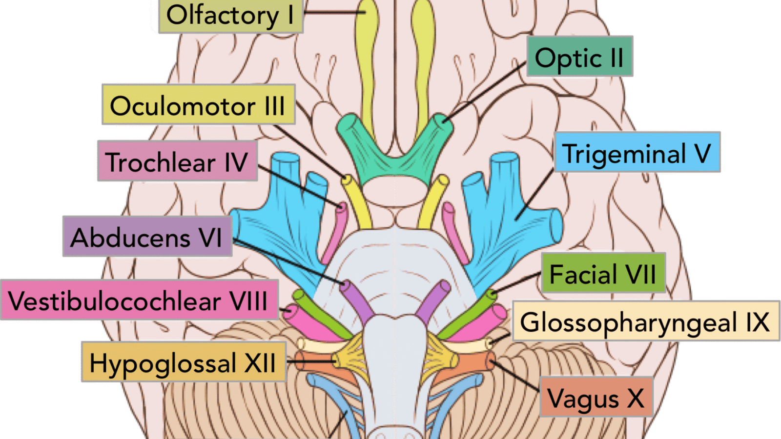
The olfactory nerve (CN I) and optic nerve (CN II) originate from the cerebrum. Cranial nerves III – XII arise from the brainstem (Figure 1).
They can arise from a specific part of the brainstem (midbrain, pons or medulla), or from a junction between two parts:
- Midbrain – the trochlear nerve (IV) comes from the posterior side of the midbrain. It has the longest intracranial length of all the cranial nerves.
- Midbrain-pontine junction – oculomotor (III).
- Pons – trigeminal (V).
- Pontine-medulla junction – abducens, facial, vestibulocochlear (VI-VIII).
- Medulla oblongata
- Posterior to the olive: glossopharyngeal, vagus, accessory (IX-XI).
- Anterior to the olive: hypoglossal (XII).
The cranial nerves are numbered by their location on the brainstem (superior to inferior, then medial to lateral) and the order of their exit from the cranium (anterior to posterior)
Let’s now explore each nerve in detail.
I. Olfactory Nerve (Cranial Nerve I)
Type: Sensory
Anatomy: Arises from the olfactory bulb and passes through the cribriform plate of the ethmoid bone.
Function: Transmits smell sensations from the nasal cavity to the brain.
Clinical Note: Damage can result in anosmia (loss of smell), which may be caused by head injury or neurodegenerative diseases.
II. Optic Nerve (Cranial Nerve II)
Type: Sensory
Anatomy: Originates from the retina and crosses at the optic chiasm before continuing to the occipital lobe.
Function: Carries visual information from the eyes to the brain.
Clinical Note: Lesions can lead to visual field defects, including blindness in one or both eyes.
III. Oculomotor Nerve (Cranial Nerve III)
Type: Motor
Anatomy: Emerges from the midbrain and innervates most of the eye muscles.
Function: Controls eyeball movement, eyelid elevation, and pupil constriction.
Clinical Note: Injury may result in ptosis (drooping eyelid), dilated pupil, or double vision.
IV. Trochlear Nerve (Cranial Nerve IV)
Type: Motor
Anatomy: Originates from the dorsal midbrain and innervates the superior oblique muscle.
Function: Assists in downward and lateral eye movement.
Clinical Note: Damage can cause vertical diplopia (double vision when looking down).
V. Trigeminal Nerve (Cranial Nerve V)
Type: Mixed
Anatomy: Divided into three branches—ophthalmic (V1), maxillary (V2), and mandibular (V3).
Function:
Sensory: Facial sensation (touch, pain, temperature)
Motor: Controls chewing muscles
Clinical Note: Can be affected in trigeminal neuralgia, a chronic pain condition causing extreme facial pain
VI. Abducens Nerve (Cranial Nerve VI)
Type: Motor
Anatomy: Arises from the pons and controls the lateral rectus muscle.
Function: Moves the eye laterally (abduction).
Clinical Note: Lesions may result in inward eye deviation and double vision.
VII. Facial Nerve (Cranial Nerve VII)
Type: Mixed
Anatomy: Travels through the internal acoustic meatus and facial canal before branching across the face.
Function:
Motor: Facial expressions
Sensory: Taste (anterior two-thirds of tongue)
Parasympathetic: Lacrimal and salivary glands
Clinical Note: Bell’s palsy involves facial nerve inflammation, causing facial droop on one side.
VIII. Vestibulocochlear Nerve (Cranial Nerve VIII)
Type: Sensory
Anatomy: Emerges from the inner ear, divided into vestibular (balance) and cochlear (hearing) branches.
Function:
Vestibular: Balance and spatial orientation
Cochlear: Hearing
Clinical Note: Lesions may cause vertigo, hearing loss, or tinnitus
IX. Glossopharyngeal Nerve (Cranial Nerve IX)
Type: Mixed
Anatomy: Originates from the medulla and exits the skull via the jugular foramen.
Function:
Motor: Swallowing
Sensory: Taste (posterior one-third of tongue), monitors blood pressure via the carotid sinus
Parasympathetic: Parotid salivary gland
Clinical Note: Dysfunction can lead to difficulty swallowing and loss of taste.
X. Vagus Nerve (Cranial Nerve X)
Type: Mixed
Anatomy: The longest cranial nerve, extending from the medulla to the thorax and abdomen.
Function:
Motor: Speech, swallowing
Sensory: Taste and visceral sensation
Parasympathetic: Regulates heart rate, digestion, and respiratory rate
Clinical Note: A critical nerve in the autonomic nervous system, vagus nerve damage can lead to heart rhythm problems, digestive issues, or voice changes.
XI. Accessory Nerve (Cranial Nerve XI)
Type: Motor
Anatomy: Arises from both the medulla and cervical spinal cord.
Function: Controls the sternocleidomastoid and trapezius muscles for head and shoulder movement.
Clinical Note: Injury can cause shoulder droop and difficulty turning the head.
XII. Hypoglossal Nerve (Cranial Nerve XII)
Type: Motor
Anatomy: Originates from the medulla and travels to the tongue.
Function: Controls tongue movement for speech and swallowing.
Clinical Note: Lesions cause tongue deviation toward the affected side and difficulty speaking.
How to Remember the Cranial Nerves
Mnemonics for Names:
“Oh Oh Oh To Touch And Feel Very Green Vegetables AH!”
Olfactory, Optic, Oculomotor, Trochlear, Trigeminal, Abducens, Facial, Vestibulocochlear, Glossopharyngeal, Vagus, Accessory, Hypoglossal
Mnemonics for Function (Sensory, Motor, Both):
“Some Say Marry Money, But My Brother Says Big Brains Matter Most”
S: Sensory | M: Motor | B: Both
These mnemonics are widely used by students and healthcare professionals to retain the order and function of cranial nerves.
Clinical Significance of Cranial Nerves
Cranial nerves are commonly assessed in neurological exams. Abnormalities in function can indicate tumors, strokes, infections, or traumatic injuries. Common signs include:
Loss of smell or taste
Visual disturbances
Facial muscle weakness
Swallowing or speech difficulties
Balance issues and vertigo
Early detection of cranial nerve dysfunction is vital for diagnosis and treatment of underlying conditions.
Cranial Nerve Disorders
Some of the most common cranial nerve-related disorders include:
Bell’s Palsy (Facial Nerve)
Trigeminal Neuralgia (Trigeminal Nerve)
Vestibular Neuritis (Vestibulocochlear Nerve)
Glossopharyngeal Neuralgia
Oculomotor Palsy
Proper imaging, physical examination, and in some cases, nerve conduction studies help in identifying these conditions.
Summary: Structure and Function of the Human Cranial Nerves
The anatomy and physiology of cranial nerves play a central role in our sensory perception, motor control, and autonomic functions. They are structurally complex and functionally diverse, making them essential for nearly every aspect of daily life—from blinking and smiling to tasting and breathing.
Understanding cranial nerves is crucial not just for students and medical professionals, but for anyone interested in how the human body works.
—Frequently Asked Questions (FAQs)
1. Which cranial nerve is the longest?
The vagus nerve (CN X) is the longest, extending from the brainstem down to the abdomen.
2. Which cranial nerves are purely sensory?
CN I (Olfactory), CN II (Optic), and CN VIII (Vestibulocochlear) are purely sensory.
3. Which cranial nerve is responsible for facial expressions?
The facial nerve (CN VII) controls all muscles involved in facial expressions.
4. How many cranial nerves are there in total?
There are 12 pairs, totaling 24 cranial nerves.
5. Can cranial nerve damage be repaired?
Some cranial nerve damage may recover over time, especially if caused by inflammation. However, severe or complete transection may lead to permanent dysfunction.
Final Thoughts
The 12 cranial nerves are the highways of communication between the brain and the body. Their anatomical paths and physiological roles are crucial to many life-sustaining processes. Whether you’re studying for an exam or seeking to understand the human body better, mastering these nerves is foundational to any exploration of the nervous system.
By understanding the anatomy and function of each cranial nerve, you’re not just memorizing facts—you’re uncovering the neural blueprint of human life.

